DISCOVER
OcuTap
TM
Investigational device. Limited by Federal law to investigational use.
Clinical Need: Addressing Challenges in Ocular Health

Over 40% of ocular inflammation cases are linked to viral infections, many of which go undetected. Without timely treatment, these infections can lead to early cataracts, scar tissue, chronic retinal swelling, and even permanent vision loss from glaucoma.
Managing ocular surface health requires precise data. OcuTap is being developed to assist clinicians by standardizing the collection of micro-volumes of ocular fluid, aiming to reduce fluid loss and minimize ambient contamination during sampling.
The Goal: Streamlining Eye Fluid Collection
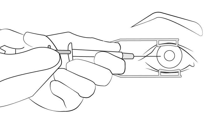
Current standard of care
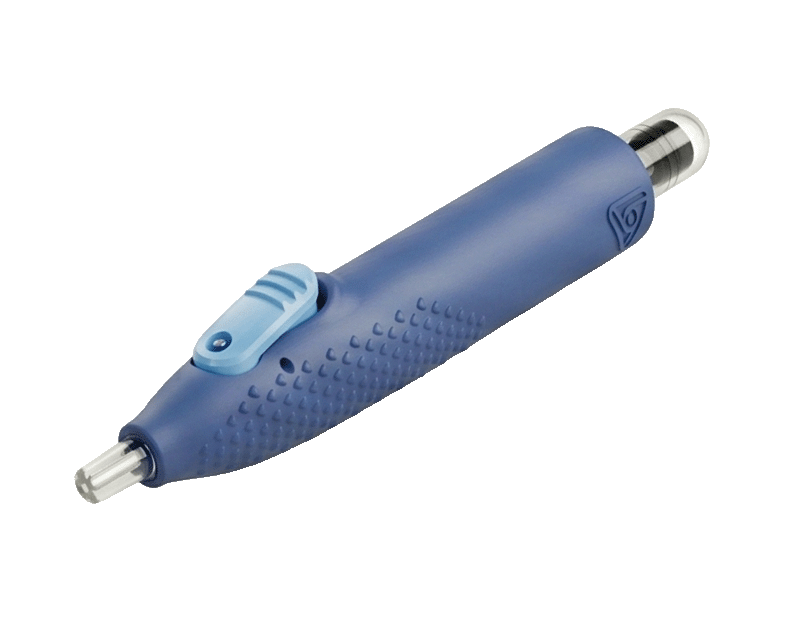
OcuTap device

Traditional eye fluid collection uses a 30-gauge needle and a 1-mL syringe, requiring extreme precision to avoid complications like iris bleeding or vision loss. The needle is inserted between the cornea and sclera to access the aqueous humor, a critical step in diagnosing infections and other eye conditions.
Our targeted design focuses on a controlled, micro-precision tip architecture. This system is intended to capture localized fluid samples quickly, mitigating common collection barriers in clinical environments.
Differentiators: OcuTap Design Objectives
Our novel technology has several key differentiators that set it apart from the current standard of care.

Single-Handed Operation
Enhances physician control and ensures a safer procedure
Minimized Sample Loss
Engineered for localized contact to capture necessary sample volumes cleanly.

Controlled Fluid Withdrawal
Designed with a smooth, micro-scale interface intended to support patient comfort during collection.

Streamlined Workflow
Built to complement standard clinical sampling steps without requiring intensive retraining.
Features: Designed for Efficiency and Safety
OcuTap is designed with both the user and patient in mind. Its ergonomic, one-handed operation simplifies fluid collection and streamlines sample preparation for shipping, reducing the risk of fluid loss and saving valuable time.
Instructions for use
Watch the video demonstration of the OcuTap procedure below, followed by detailed, step-by-step instructions.
The following protocol outlines the intended engineering functionality of the OcuTap™ device. These instructions are provided for informational and investigational purposes only, pending official regulatory clearance.
1. Attach needle. Securely attach a sterile needle (not included) to the device. Do not remove the needle cap at this time.
2. Prime the device. Pull back the collection tube until it reaches an extended position and clicks into place.

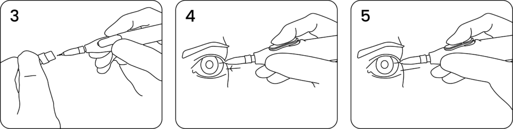
3. Uncap needle. Carefully remove the protective cap from the needle, ensuring the tip remains sterile and does not come into contact with any surrounding surfaces prior to insertion.
4. Insert & collect fluid. Insert the device into the eye at the limbus, strictly following standard anterior chamber paracentesis procedures. Once safely positioned, depress the central button to open the valve and begin the collection of fluid. If necessary at any point, release the button to immediately stop the fluid flow.
5. Withdraw device. IOnce the collection is finished, release the button and carefully withdraw the device from the eye.
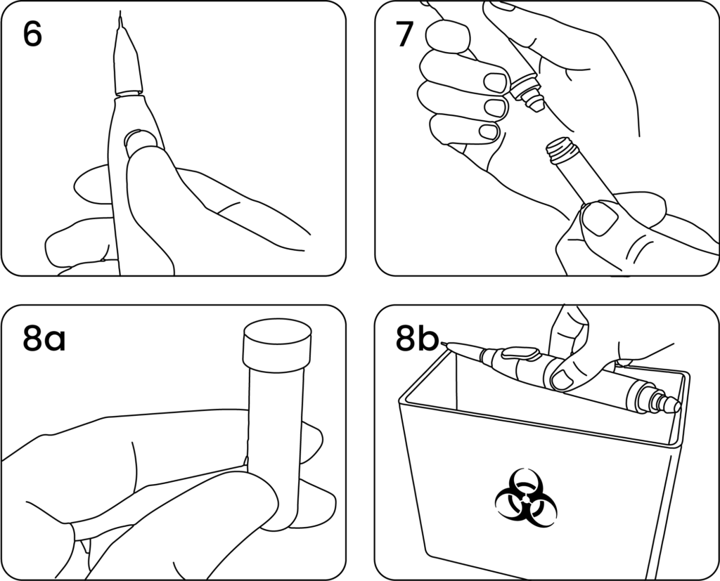
6. Clear internal pathway. Give the button a final push to move any remaining fluid from the internal pathway into the collection tube.
7. Remove collection tube. Twist the collection tube to detach and remove it from the primary device.
8. Dispose and secure. Cap the barrel, and safely discard the syringe body in a sharps container. Place a cap securely on the collection tube for shipping and analysis.